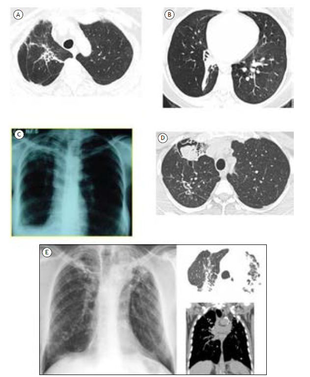
Figure 1. Radiological patterns of post-tuberculosis lung disease. In A, a chest CT scan showing irregular dense opacity with bronchiectasis from the hilum to the right lung apex associated with apical pleural thickening, elevation of the hilum, and volumetric reduction on this side. In B, a chest CT scan showing consolidative opacity with a fibroatelectatic appearance in the right lower lobe, predominantly affecting the anterior, lateral, and posterior basal segments, determining its volumetric reduction and highlighting the associated bronchiectasis in the anterior basal segment. Cylindrical bronchiolectasis in the upper segment of the right lower lobe. In C, a chest X-ray showing fibrotic opacity in the right upper lobe with pleural thickening and volumetric reduction of the right lung, leading to elevation of the right phrenic dome. In D, a chest CT scan showing a residual excavated lesion in the anterior segment of the right upper lobe filled with mobile contents upon change of decubitus, corroborating repercussions of superimposed saprophytic fungal infectious involvement. In E, imaging scans showing confluent laminar atelectasis, volumetric loss, and architectural distortion in the upper lobes, with intervening calcifications, favoring chronic/residual changes.

