Figure 2.
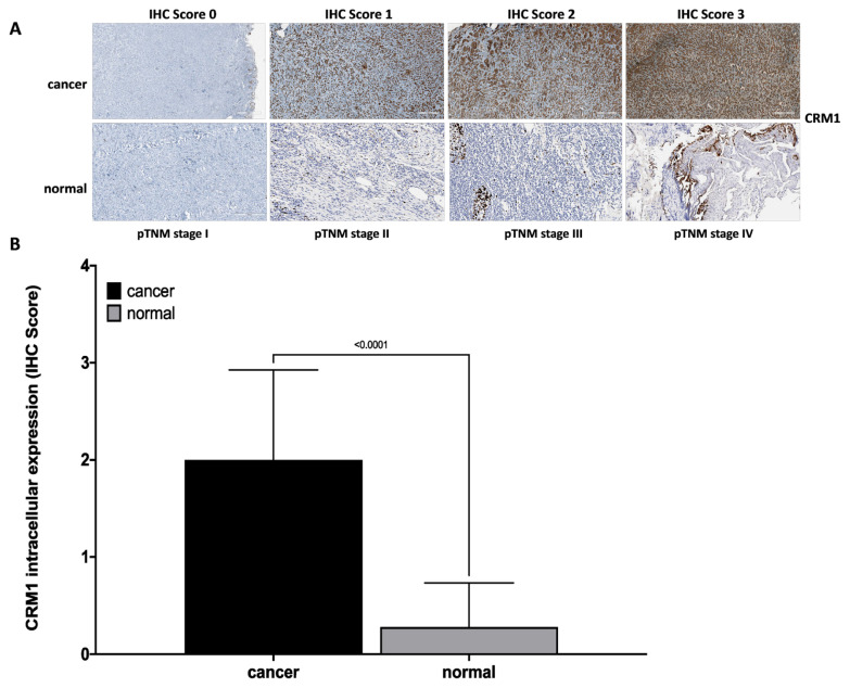
IHC data showing intracellular expression of CRM1 in matched tumor and normal tissue sections from laryngeal cancer patients. (A) Representative microscopic images of CRM1 staining scored by IHC method from laryngeal tumor and normal tissue sections at pTNM stage I, II, III, and VI (100× magnification, scale bar = 100 μm). CRM1 showed intense nuclear and cytoplasmic staining in tumor tissue and weak immunoreaction in normal laryngeal epithelium. In addition, as the tumor stage increased, an increase in the IHC score for CRM1 was observed. (B) IHC score data of CRM1 in matched tumor and normal tissues from laryngeal cancer patients. Intracellular expression of CRM1 was significantly higher in tumor tissues compared to normal tissues (p < 0.0001). IHC score data was obtained from at least three independent experiments. The intensity and amount of immunoreactivity in microscopic images obtained from cells stained with CRM1 antibody in ten randomly selected fields in each preparation were evaluated using Aperio ImageScope 12.4.3 software. Student’s t-test was used for statistical analysis. IHC score: 0 (no staining), +1 (weak staining), +2 (moderate staining), +3 (strong staining). Results are expressed as mean ± SD. CRM1: the chromosome region maintenance 1 protein, IHC: Immunohistochemical, SD: Standard error.

