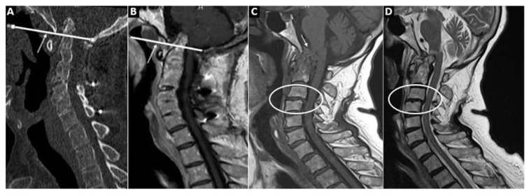
Figure 3.
A (cervical CT) and B (cervical MRI) show the Chamberlain line and the associated vertical subluxation. In C (T1WI) and D (T2WI), basilar impression with pannus tissue at the craniovertebral junction causing stenosis of the foramen magnum and compression of the brainstem are shown. In addition, subaxial subluxation between C3–4 is seen.

