
Figure 5.
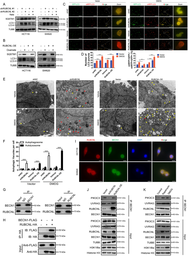
RUBCNL promoted autophagasome maturation through mediating the recruitment and function of PtdIns3K complex.
Note: (A and B) Western blot analysis of LC3-II:I and SQSTM1 expression in HCT116 and SW620 cells after RUBCNL silencing and supplemented with 5 mM Nala (A) or with or without RUBCNL overexpression treated with 8 mM oxamate (B). (C and D) mRFP-GFP-LC3 was expressed in HCT116 cells transfected with shRUBCNL or RUBCNL-expressing plasmid, respectively. Cells were treated with DMSO (left panel) or 2 mM DMOG (right panel) for 6 h. The number of LC3 puncta was analyzed by fluorescence microscope (C). GFP-negative mRFP-positive (GFP-mRFP+) puncta, which indicate autolysosomes, were quantified and are summarized in (D) (scale bar: 100 μm). *P ≤0.05, **P ≤0.01, ***P ≤0.001. (E and F) HCT116 cells transfected with shRUBCNL or RUBCNL-expressing plasmid, respectively, were treated with DMSO or 2 mM DMOG for 6 h and then analyzed by transmission electron microscopy (E) (scale bar: 0.5 μm). Arrows indicate autophagic vacuole (red arrows, autophagosome; yellow arrows, autolysosome). The autophagic vacuole per cross-sectioned cell under EM was calculated and is summarized in (F). ***P ≤0.001. G. Endogenous interaction between BECN1 and RUBCNL was determined using co-IP with anti-BECN1 or anti-RUBCNL antibodies in HCT116 cells. (H) Exogenous interaction between BECN1 and RUBCNL was determined using co-IP with anti-FLAG or anti-HA antibodies in HEK 293T cells co-transfected with BECN1-FLAG and RUBCNL-HA. (I) Immunofluorescence staining showing colocalization of endogenous RUBCNL (red) and BECN1 (green) in HCT116 and SW620 cells. The nucleus is labeled by DAPI (blue) (scale bar: 20 μm). (J) Endogenous PtdIns3K complexes were IP from cells with Vector, shRUBCNL, or RUBCNL overexpression using anti-BECN1 antibody, and then they were analyzed by western blot. (K) Endogenous PtdIns3K complexes were IP from control cells and cells treated with 200 nM rapamycin or 2 mM DMOG using anti-BECN1 antibody, and they were analyzed by western blot.