
Figure 4.
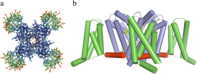
Structure of the bacterial sodium channel NaVAb. a) top view of NaVAb channels colored according to crystallographic temperature factors of the main-chain (blue <50 Å2 to red > 150 Å2). The four pore modules in the center are rigid in the crystal structure and therefore are blue. The four voltage-sensing modules surround the pore and are more mobile, as illustrated by warmer colors. b) side view of NaVAb. Voltage sensing module (S1-S4), green; pore module (S5, S6, and P loop), blue; selectivity filter, yellow; S4-S5 linker, red. Adapted from Payandeh et al.,2011 [97].