
Figure 5.
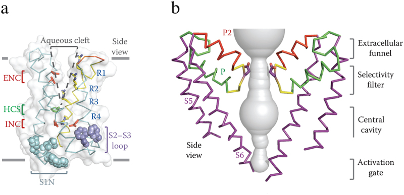
Architecture of the NaVAb voltage sensing module and pore module. a) side view of the voltage-sensing module of NaVAb illustrating the conformations of the S1-S4 helices and the size of the extracellular aqueous cleft with the R1-R4 gating charges (blue), extracellular negative cluster (ENC, red), intracellular negative cluster (INC, red), and hydrophobic constriction site (HCS, green). b) side view of the pore-forming module of two subunits of NaVAb. S5 and S6 transmembrane helices, purple: P helix loop, green; P2 helix green; water-filled space revealed by MOLE, gray. Note that the two S6 segments are crossed at their intracellular ends, forming the closed conformation of the activation gate. Adapted from Payandeh et al., 2011 [97].