
Figure 8.
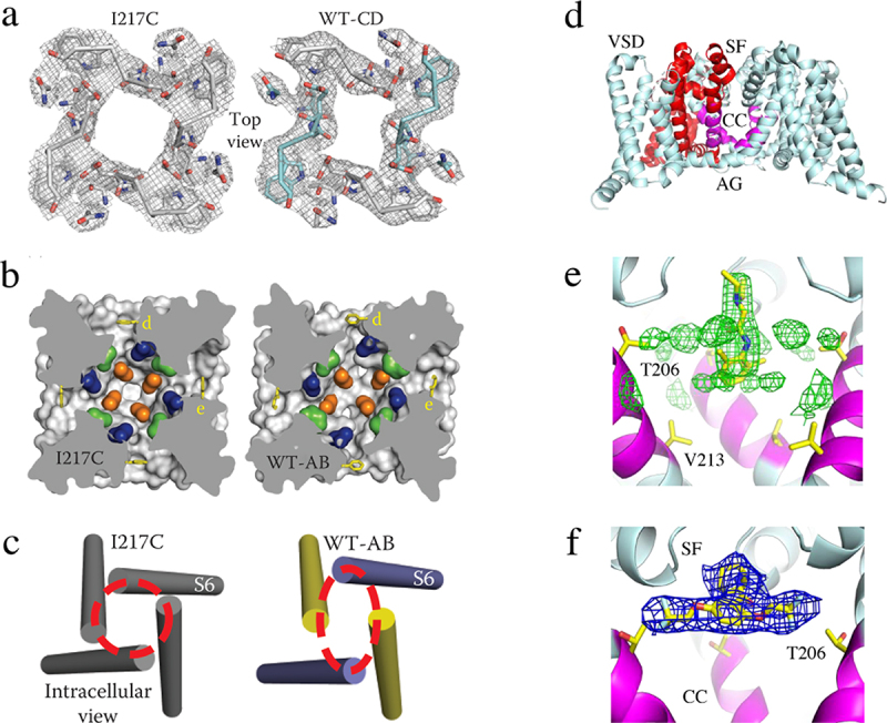
Slow inactivation and drug block of Na+ channels. a) top view of the collapse of the pore during slow inactivation of NaVAb. Two S6 segments move inward the central axis of the pore and two move outward to produce an asymmetric, partially collapsed conformation. The selectivity filter structure has changed from nearly square in the pre-open state of NaVAb/I217C to a partially collapsed parallelogram in the inactivated state of NaVAb/WT-CD. b) the central cavity is partially collapsed. c) the activation gate is tightly closed, but collapsed into a two-fold symmetric conformation. Adapted from Payandeh et al, 2012 [102]. d) structure of NaVAb with the selectivity filter (SF), central cavity (CC), and activation gate (AG) highlighted. e) lidocaine bound in the central cavity at the base of the selectivity filter. f) flecainide bound at the local anesthetic/antiarrhythmic receptor site in the central cavity at the base of the selectivity Filter.