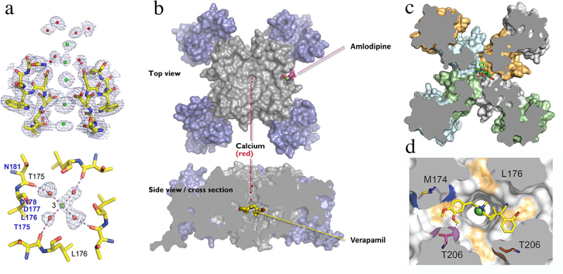
Figure 9.

Calcium and drugs binding to the pore module of CaVAb. a) side view of the ion selectivity filter of CaVAb from X-ray crystallography. a) the ion selectivity filter of CaVAb at high resolution. Green balls, calcium ions; red balls, water; mesh, electron density. Top. side view; bottom, bottom view illustrating a single calcium ion with a square array of four waters of hydration bound. Adapted from Tang et al., 2014 [113]. b) structure of CaVAb in top and side views by X-ray crystallography. Blue, voltage sensor; gray, pore module. Bound amlodipine and verapamil, yellow sticks. c) top view of a cross-section of CaVAb with diltiazem bound in its receptor site as indicated (green sticks). Adapted from Tang et al., 2018 [138]. d) bottom X-ray crystallographic view of a cross-section in high resolution with verapamil (yellow sticks) bound in its receptor site and a calcium ion (green) bound in the pore. Adapted from Tang et al., 2014 [113].
