
Figure 11.
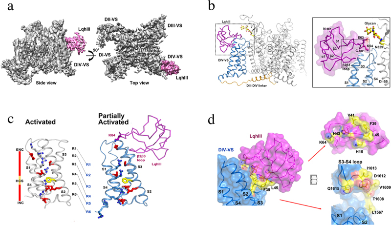
Receptor site for the deathstalker scorpion toxin LqhIII on NaV1.5. a) spacefilling side view and top view of NaV1.5 (gray) with LqhIII bound (purple). b) close-up view of bound LqhIII (purple) to its receptor site on the NaV1.5 backbone structure. Inset. higher resolution image of the interface between LqhIII and NaV1.5. c) superposition of the voltage sensor in the unmodified activated state and the toxin-modified partially activated state. d) mutational map of the interface residues in the LqhIII receptor site illustrated with “open book” format. LqhIII, purple; NaV1.5, blue. Amino acid residues highlighted in yellow and orange are required for high-affinity binding. Adapted from Jiang et al., 2021 [168].