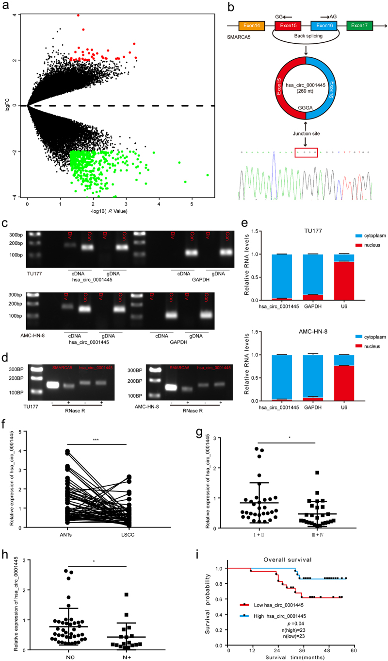
Figure 1.

Identification and characteristics of hsa_circ_0001445 in LSCC and the expression of hsa_circ_0001445 in LSCC patients and its clinical significance.
(a) Volcano plot of the differentially expressed circRNAs in five pairs of LSCC tissues and matched adjacent non-tumor tissues. The red dots represent higher expression levels, while the green dots represent lower expression levels. (b) Schematic illustration showed the circularization of SMARCA5 exons 15 and 16 to form hsa_circ_0001445. Sanger sequencing following RT-PCR was used to show the “head-to-tail” splicing of hsa_circ_0001445. (c) Hsa_circ_0001445 expression in TU177 and AMC-HN-8 cells verified by RT-PCR. Agarose gel electrophoresis showed that divergent primers amplified hsa_circ_0001445 in cDNA but not in gDNA. GAPDH served as a negative control. (d) Validation of hsa_circ_0001445 stability by RNase R treatment and RT-PCR assay. (e) Hsa_circ_0001445 abundance in nuclear and cytoplasmic fractions of TU177 and AMC-HN-8 cells was evaluated by qRT-PCR. GAPDH acted as a positive control of RNA distributed in the cytoplasm, and U6 RNA acted as a positive control of RNA distributed in the nucleus. (f) Expression levels of hsa_circ_0001445 in 57 paired LSCC tissues were determined by qRT-PCR. (g-h) The expression of hsa_circ_0001445 in different groups was evaluated according to the clinical features (n (clinical stageI+II) = 31, n (clinical stageIII+IV) = 26; n(N0) = 40, n(N+) = 17). I Kaplan-Meier analysis of the correlation between hsa_circ_0001445 expression and overall survival of 46 LSCC patients. Data represent means ± SD of three independent experiments. *P< 0.05, **P< 0.01, ***P< 0.001. Div, divergent primer; Con, convergent primer; ANTs, adjacent non-tumor tissues; N0, patients without cervical lymph node metastasis; N+, patients with cervical lymph node metastasis.
