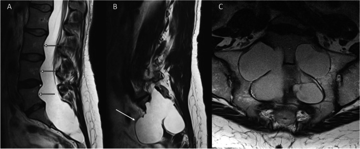
Fig. 2.
T2-weighted sagittal and axial images of the lumbosacral spine which demonstrate dural ectasia in a patient with Marfan’s syndrome. A, B Sagittal images illustrate posterior vertebral body scalloping (arrows) and anterior sacral meningoceles (arrows). C Axial image shows marked enlargement of the neural foramina associated with dural ectasia. Dural ectasia is a feature of other hereditary connective tissue disorders including Loeys-Dietz and Ehlers-Danlos syndrome

