Fig. 4.
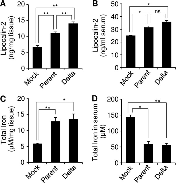
Effect of SARS-CoV-2 infection on the iron concentration in a lung tumor xenograft mouse model. Calu-3 cells were subcutaneously injected into the right flank of NRGA mice (n = 3/group). Parental SARS-CoV-2 or SARS-CoV-2 Delta was intratumorally infected. (A, C) Supernatants of tumor homogenates were collected at 15 DPI, and then, the Lpocalin-2 concentration was determined by ELISA (A). The total iron concentration in the tissues was determined using the supernatants of the tumor homogenates by the Iron Colorimetric assay kit (C). (B, D) Sera were collected at 15 DPI in a lung tumor xenograft mouse model, and then, the lipocalin-2 concentration was determined by ELISA (B). The total iron concentration in sera was determined by the Iron Colorimetric assay kit (D). Values are the mean ± SEM. *P < 0.05, **P < 0.01 vs. mock control.

