Figure 3.
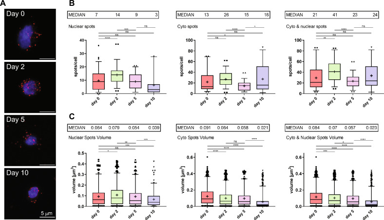
HBL1 nuclear and cytoplasmic localization during hiPSC differentiation toward cardiomyocytes by RNA FISH. Human induced pluripotent stem cells were differentiated into cardiomyocytes over 10 days. On days 0, 2, 5, and 10, cells were collected and subjected to RNA FISH targeting HBL1 lncRNA and analyzed using confocal microscopy. (A) Representative images of cells on days 0, 2, 5, and 10 as maximal projections reconstructed from confocal image stacks. HBL1 RNA is shown in red, and DAPI-stained nuclei are shown in blue. Scale bar = 5 µm. The number (B) and volume [µm3] (C) of nuclear, cytoplasmic, and total (nuclear + cytoplasmic) HBL1 lncRNA foci. Images of single cells were acquired with a confocal microscope, and HBL1 RNA foci were quantified inside DAPI-stained nuclei and in the cytoplasm in z-stacks from 10 images/sample in three independent differentiations (n = 30). The results are presented as the mean values (depicted as “ + ”) ± s.d. Median values are shown as bars and indicated above each graph. Statistics were performed using the Welch two-sample t-test. Statistical comparisons are indicated if p < 0.05.

