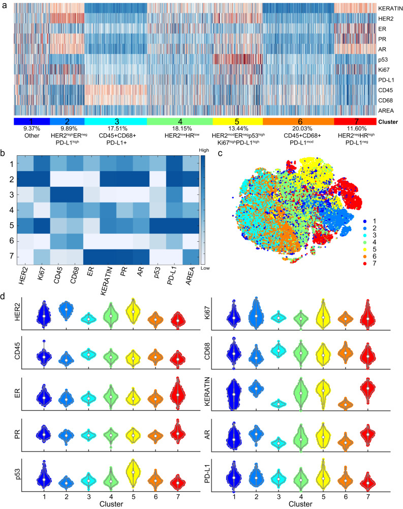
Fig. 5. CyCIF single-cell analysis of HER2+ breast cancer reveals tumor populations with heterogenous HER2 expression.
Following the selection of qualified ER, PR, HER2, AR, and p53 CyCIF antibodies, the expression of selected antibodies was evaluated at a single-cell level in 567 HER2+ invasive breast cancer samples, representing 189 patients. a Single-cell clustering is shown, and b median expression for each antigen across each cluster is shown. Relative expression of HER2 is designated as high, moderate (mod), low, and negative among clusters 2, 5, 4, and 7, respectively. Tumor clusters are defined as: cluster 2 (HER2highERnegPRposARposPD-L1highKi67pos), cluster 5 (HER2modERnegPRposARposp53highPD-L1posKi67pos), and cluster 4 (HER2lowERposPRlowARpos)). One cluster was HER2-negative (cluster 7 (HER2negERhighPRhighARhigh)). Cluster 3 and 6 represent immune/stromal populations as characterized by the expression of the leukocyte marker CD45. Cluster 1 has heterogeneous expression of most proteins and, therefore, did not form a distinct population of cells. Area in A and B refers to the nuclear area of segmented cells. c The 7 cell clusters are visualized using t-SNE. d Volcano plots of expression of each marker by cluster.

