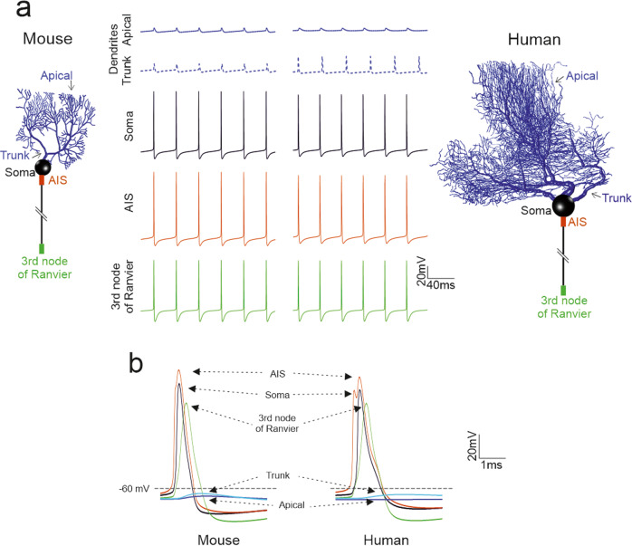
Fig. 4. Spike propagation in PC models.
(a) In all PC models, spikes are generated in the AIS and then propagate actively in the axon and passively in the dendrite, where they undergo a marked decrement and slowdown. Traces show the spontaneous spike discharge in different model compartments. The corresponding PC model morphologies are shown on the side (with the colors indicating the recording site). (b) Spikes taken from the same simulations as in A are overlay for AIS, soma, axon, aspiny, and spiny dendrites. Note that the AIS spike precedes those in any other compartment (same colors as in a).

