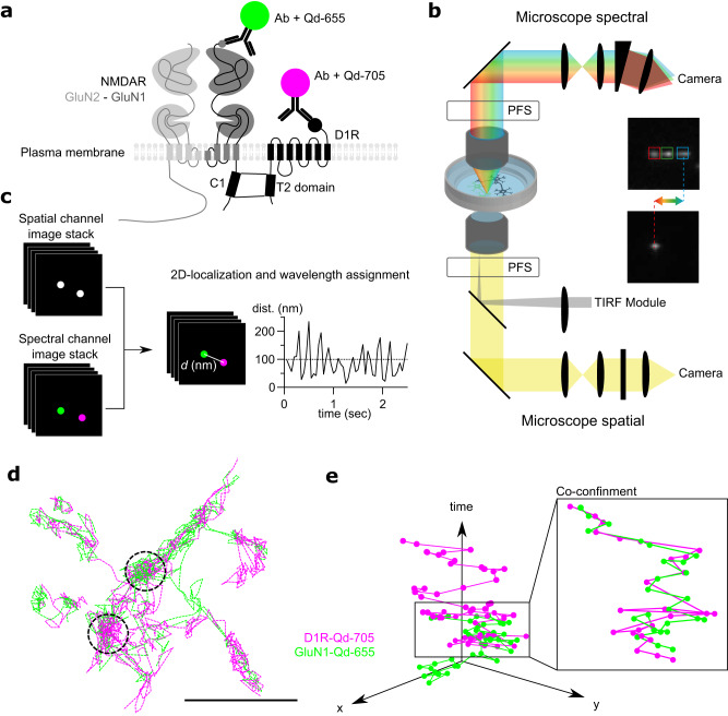
Fig. 1. Multidimensional spectral single molecule localization microscopy (MS-SMLM) principle.
a Experimental design of the single Qd tracking. Receptors were labeled with antibodies directed against extracellular tags. Qd-655 and Qd-705 were used to distinguish receptor types. The interaction between receptors occurs intracellularly at the T2 domain (C1 cassette of the GluN1 subunit). b Microscopy setting to perform MS-SMLM using two microscopes and cameras. PFS: perfect focus system. c SM-SMLM principle. d Representative reconstruction of GuN1-NMDAR and D1R surface diffusion, scale bar, 1 µm. e Example trajectories of one GluN1-NMDAR and one D1R laterally diffusing (x, y) onto the neuronal surface over time.

