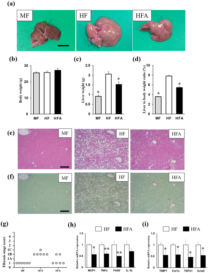
Figure 1.
C57BL6/J mice were fed a regular diet (MF), or choline-deficient, l-amino acid-defined, high-fat diet (HF) with/without 5% ASHE (HFA) for 6 weeks. (a) Macroscopic images of the liver in each diet-fed mouse group. Scale bars, 10 mm. (b) Total body weight. (c) Liver weight. (d) Liver to body weight ratio (% body weight). *P < 0.001, statistical significance when compared to HF-fed mice. (e) Microscopic images of the liver stained by hematoxylin–eosin staining. (f) Microscopic images of the liver stained by Sirius Red-Fast Green staining. Scale bars, 200 µm. (g) Fibrosis stage score. (h) Gene expression of inflammation-related genes. (i) Gene expression of fibrosis-related genes. Data represent the mean ± SD (n = 6). *P < 0.05, **P < 0.01, statistical significance compared to HF-fed mice.

