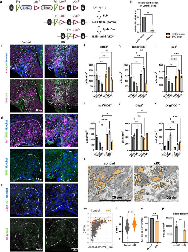
Fig. 1. IL4I1 deletion in myeloid cells results in increased myeloid cell-driven inflammation and decreased remyelination.
a Illustration of conditional knockout IL4I1 in myeloid cells and experimental controls. b RT-qPCR of Il4i1 mRNA levels in MACS-isolated CD11b+ myeloid cells from the lesions of IL4I1-cKO mice and controls (n = 3, each n was obtained from 3–5 mice, unpaired t test). Representative images for immunostaining of mouse lesions for CD68+pS6+ myeloid cells at 10 dpl (c), Iba1+iNOS+ inflammatory myeloid cells at 20 dpl (d), and Olig2+CC1+ mature oligodendrocytes at 20 dpl (e). f–k Quantification of immunostaining cell counts in the lesions of IL4I1-cKO mice and littermate controls at 5, 10, 20 dpl (n = 3–5 mice, two-way ANOVA). l Representative images for SEM of spinal cord lesions of IL4I1-cKO mice and littermate controls at 20 dpl. m, n Quantification of g-ratio from SEM images (n = 2 mice for both groups, 195–296 axons from each n, unpaired t test). o Quantification of percentage of axons remyelinated (n = 2, unpaired t test). p Quantification of axonal density within the demyelinated lesions of controls and IL4I1-cKO mice. Bars represent the means with SEM. Each point represents an individual value. *P < 0.05, **P < 0.01, ***P < 0.001, ****P < 0.0001, ns = not significant.

