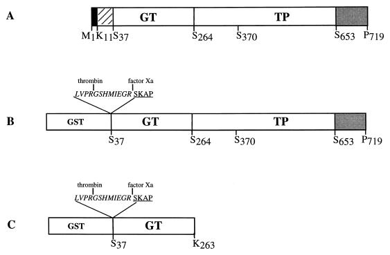
FIG. 2.
Schematic diagrams of the PBP 1a-derived constructs. (A) Organization of the native PBP 1a protein. Filled, hatched, and shaded boxes indicate the N-terminal cytoplasmic region, the membrane anchor, and the Ser- and Asn-rich C-terminal extension, respectively. (B) Construction of the GST-PBP 1a* fusion protein. (C) Construction of the GST-GT fusion protein. The active-site serine 370 is indicated. Peptide at the GST-GT junctions includes sequences specific for thrombin and factor Xa (italic characters) and N-terminal amino acids of GT (underlined characters).

