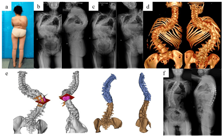
Figure 2. Spinal deformity.
This is an open-source image: (a) kyphoscoliosis deformity, (b) X-ray of the entire spine in a standing position before surgery, (c) X-ray of the entire spine while bending, (d) three-dimensional reconstruction of the complete spinal computed tomography scan, (e) simulation of vertebral column resection osteotomy, and (f) after surgery, an X-ray of the entire spine in a standing position reveals significant improvement in the deformity [20].

