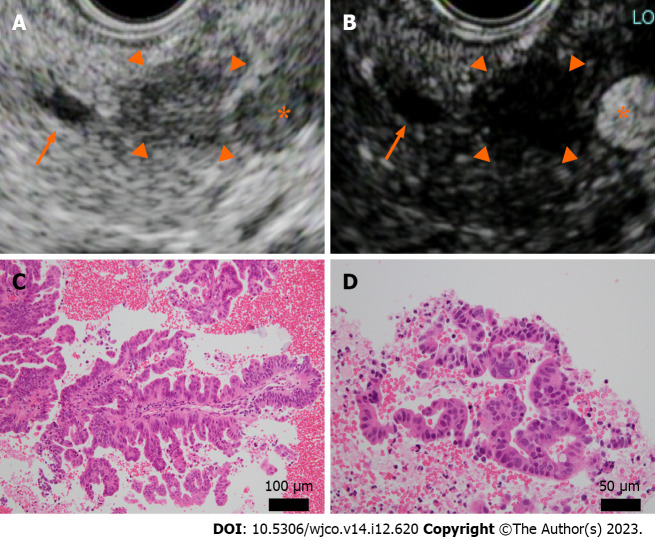
Figure 2.
Endoscopic ultrasound and histopathology (hematoxylin-eosin staining) at the time of endoscopic ultrasound-guided fine needle aspiration. A and B: A well-defined hypoechoic mass 10 mm in size was observed in the pancreatic body (A), arrowhead. The main pancreatic duct, arrow; splenic artery, asterisk. The mass was recognized as an oligo-hypoechoic mass with Sonazoid® contrast agent (B), arrowhead; C and D: Atypical epithelium with ductal papillary growth was seen. No intraductal papillary mucinous tumor-like mucus component was present. Original magnification was × 20 (C) and × 40 (D).

