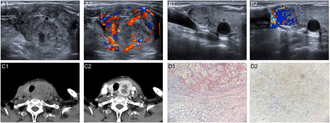
Figure 1.
A 78-year-old woman with thyroid metastasis from ccRCC. A1. Transverse sonogram showing a heterogeneous hypoechoic nodule (4.2 × 3.3 × 2.9 cm) with irregular morphology in the left lobe. A2. Color Doppler sonogram shows abundant blood flow signals in the nodule. B1. Transverse sonogram showing a hypoechoic nodule (16 × 11 mm) with regular morphology in the right lobe. B2. Color Doppler sonogram image shows abundant blood flow signals in the nodule. C1. CT showing low-density masses in both lobes of the thyroid gland with poor demarcation from adjacent muscle and soft tissue. C2. Uneven enhancement on enhancement scan. D1. Surgical pathology confirming thyroid metastasis from ccRCC (HE ×200). D2. Immunohistochemistry shows CD10 (+) (HE ×400).

 This work is licensed under a