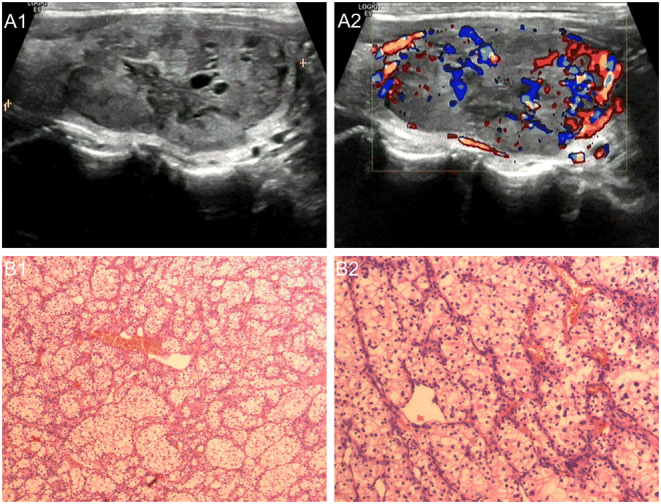
Figure 2.

A 54-year-old man with thyroid metastasis from ccRCC. A1. Transverse sonogram showing a partially cystic nodule (4.5 × 2.0 × 3.5 cm) with clear borders and regular morphology in the left lobe. A2. Color Doppler sonogram image shows abundant blood flow signals in the nodule. B1 and B2. Hematoxylin and eosin staining; Surgical pathology confirming thyroid metastasis from ccRCC.

 This work is licensed under a