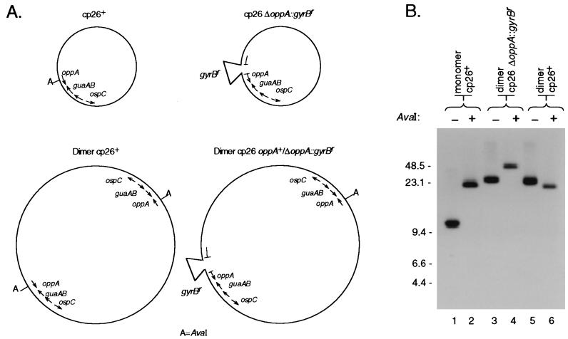
FIG. 4.
AvaI digestion patterns of various plasmid preparations. (A) Schematic showing the positions of AvaI sites on wild-type and ΔoppAIV::gyrBr cp26. (B) Southern blot analysis of uncut and AvaI-digested cp26 and cp26* from wild-type bacteria and ΔoppAIV::gyrBr mutants. Plasmid preparations from the indicated clones were electrophoresed through a 0.3% agarose gel with (+) or without (−) AvaI digestion. DNA was transferred to a nylon membrane and hybridized with a guaA probe (Table 2). Lanes: 1 and 2, B31-86; 3 and 4, B31-34; 5 and 6, B31-9. Marker sizes are in kilobases.

