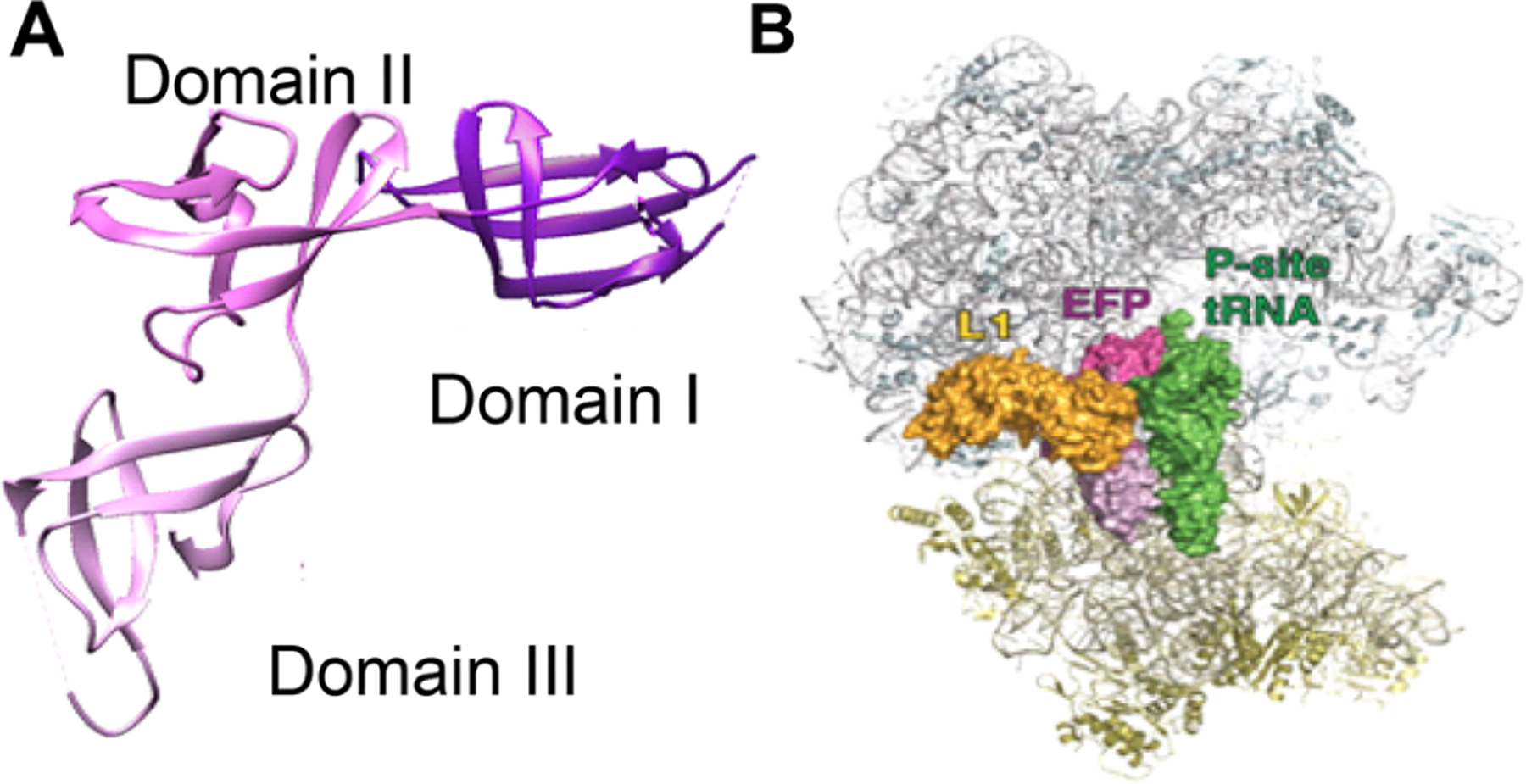
Figure 1.

Structure of Clostridium thermocellum EF-P (pdb 1YBY). (A) The three structural domains of EF-P are shown in different shades of magenta. (B) An overview of the binding of EF-P to the 70S ribosome (pdb 2j00 and 2j01; adapted from Blaha, G.; Stanley, R. E.; Steitz, T. A., Science 2009, 325, 966–9709). Reprinted with permission from AAAS. EF-P is in magenta, while the 50S subunit is colored gray, and the 30S subunit is in yellow. Additionally, ribosomal protein L1 is shown in gold, and the P-site tRNA is in green.
