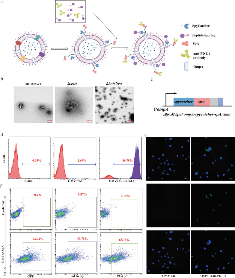
Fig. 1.
Generation and characterization of OMV-PP. a Schematic illustration of the OMV-based delivery system. The OMVs extracted from ΔlpxMΔpal strain were bound with both SpT-attached TAAs and PD-L1 antibody. b Representative TEM image of OMVs. Scale bar: 200 nm. c Sketch map of ΔlpxMΔpal ompA-spC-spA (the drawing is not to scale). d, e Examination of the ability of OMV-SpA to capture PD-L1 antibody. d) Flow cytometry analysis and e confocal microscopy images (blue, DAPI; green, FITC). Scale bar: 70 μm. f Testing the capability of OMV-SpC-SpA to capture SpT-attached mCherry, GFP, and PE-Cy7-labeled IgG antibody

