Fig. 2.
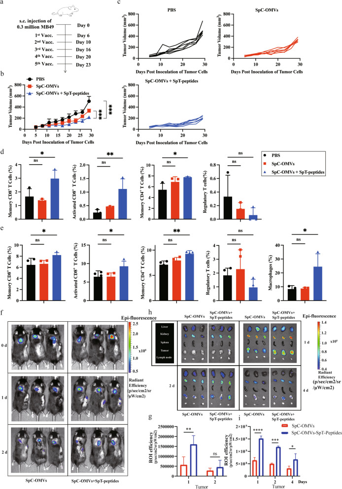
TAA-bound OMVs specifically targeted tumors and repressed bladder cancer growth. a Schematic illustration examining how OMV-P inhibits bladder cancer growth. C57BL/6 mice were s.c. injected with MB49 cells in the right flank on day 0 and s.c. immunized with PBS, OMV, or OMV-P on days 6, 10, 16, 20 and 23. b, c Antitumor effect of OMV-P. Tumor size was measured every 3 days. Quantification of the tumor size data are presented in (b, c). Experiments were repeated three times. d) Quantitative analysis of different subsets of CD3+CD45+ T cells in lymph nodes by flow cytometry (n = 3). Memory CD4+ T cells, CD4+CD44+ T cells. Memory CD8+ T cells, CD8+CD44+ T cells. Activated CD8+ T cells, CD8+Granzyme-B+ T cells. Treg, CD4+CD25+FoxP3+ T cells. e) Quantitative analysis of different subsets of immune cells in PBMCs by flow cytometry (n = 4 or 3). Memory CD4+ T cells, memory CD8+ T cells, activated CD8+ T cells, and Treg were analyzed as described above (n = 4). Macrophages (n = 3), CD45+CD11b+F4/80+ cells. f, h) Cy7-labeled OMV-P were systemically injected into C57/BL6J mice bearing MB49 tumor cells. Whole body distribution of the injected Cy7-OMV-P was observed by an in vivo imaging system on days 0, 1, and 2 after injection. Liver, kidney, spleen, tumor, and lymph node were isolated to measure the accumulation of Cy7 fluorescence in different organs. d, day. g, i Quantification of fluorescence in the tumors (in vivo or ex vivo) (n = 3). Data are shown as mean ± SD. One-way ANOVA with a Tukey multiple comparisons test. NS, no significance; *, p < 0.05; **, p < 0.01; ***, p < 0.001; ****, p < 0.0001

