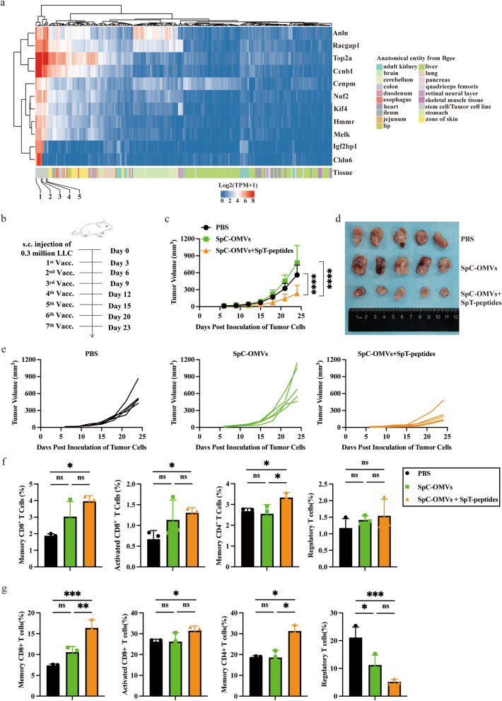
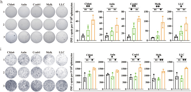
Fig. 3.
TAA-modified OMVs significantly suppressed lung cancer growth. a RNA sequencing revealed the expression profiles of 11 selected genes between mouse ESC and tumor cell lines compared to normal healthy tissues. 1. ESC (129); 2. ESC (C57BL/6); 3. Hepa1 − 6 cell; 4. LLC cell; 5. MB49 cell; b Schematic illustration examining how OMV-P inhibits lung cancer growth. C57BL/6 mice were s.c. injected with LLC cells in the right flank on day 0 and s.c. immunized on day 3, 6, 9, 12, 15, 20, and 23 for seven total vaccinations. c-e Antitumor effects of OMV-P. Tumor size was measured every 3 days. f, g Quantitative analysis of different subset of immune cells in lymph nodes f and PBMCs g by flow cytometry (n = 3). h, i Quantitative analysis of ELISPOT assay for IFNγ secretion to detect immune cell activation in splenocytes (h) and TILs (i) against selected epitopes and LLC tumor cells. 1, PBS. 2, SpC-OMVs. 3, SpC-OMVs + SpT-peptides. Data are shown as mean ± SD. One-way ANOVA with a Tukey multiple comparisons test or unpaired two-tailed Student’s t-test. NS, no significance; *, p < 0.05; **, p < 0.01; ***, p < 0.001; ****, p < 0.0001


