Fig. 6.
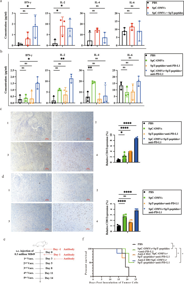
The antitumor effects of OMV-PP were T cell immunity-dependent. a, b) Multiplex cytokine assay was performed to determine the expression profiles of serum IL-2, IL-4, -IL-6, and INF-γ in lung cancer model (a) and bladder cancer model (b). c, d IHC images showing the infiltration level of CD4+ and CD8+ T cells in tumor tissues. 1, PBS. 2, SpC-OMVs. 3, SpT-peptides + anti-PD-L1. 4, SpC-OMVs + SpT-peptides + anti-PD-L1. Scale bar: 100 μm. Quantitative analysis on the right. Data are shown as mean ± SD. e Schematic illustration determining whether CD4+ and CD8+ T cells are integral for the antitumor effects of OMV-PP. C57BL/6 mice were s.c. inoculated with MB49 cells in the right flank on day 0 and s.c. immunized on day 3, 5, 8, 11 and 14 for five total vaccinations. From day -1, 1, and 3 after tumor inoculation, 10 mg kg−1 of CD8+ T cell depletion antibody and 10 mg kg−1 of CD4+ T cell depletion antibody were injected intraperitoneally in mice. f Survival time of MB49 tumor-bearing mice were measured. One-way ANOVA with a Tukey multiple comparisons test or unpaired two-tailed Student’s t-test. NS no significance; *, p < 0.05; **, p < 0.01; ***, p < 0.001; ****, p < 0.0001

