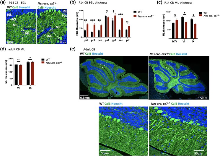
Fig. 2.
Cerebellar pathology in Nes-cre, ex7−/− mutant mice. a) Representative image of the base of the fissures in P14 WT and Nes-cre, ex7−/− cerebellum; white boxes enclose the external granule layer (EGL) at the base of the fissures; dashed lines show the thickness of the molecular layer (ML); PCL, Purkinje cell layer; IGL, inner granule layer. b) Quantification of EGL thickness at the base of different fissures in P14 WT and Nes-cre, ex7−/− cerebellum vermis. Three measurements were averaged for each section examined; N = 3–6 matching sections from 3 mice for each genotype. pri, primary fissure; pcf, preculminate fissure; pce, precentral fissure; psf, posterior superior fissure; ppf, prepyramidal fissure; sec, secondary fissure; plf, posterolateral fissure; c) quantification of ML thickness in P14 WT and Nes-cre, ex7−/− vermis. Three measurements were averaged for each location examined on per section; N = 5–6 matching sections from 3 mice for each genotype. IV/V, VI, and IX indicate lobules examined. d) Quantification of ML thickness in adult WT and Nes-cre, ex7−/− vermis. Three measurements were averaged for each section examined; N = 6–8 matching sections from 3 mice for each genotype. VI and IX indicate lobules examined. e) Representative image of CB in adult WT and Nes-cre, ex7−/− mice (upper panels); dashed boxes circle zoomed in view of lobule VI (lower panels); 2-tailed t-test was used to test statistical significance, *P < 0.05, **P < 0.01, ****P < 0.0001, ns = not significant.

