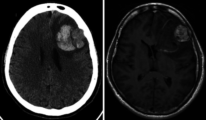
FIG. 1.

Left: Axial noncontrast computed tomography (CT) demonstrating a left frontal lobe hematoma with surrounding vasogenic edema and mild midline shift, as well as an adjacent peripherally centered, 2.9-cm, hyperdense mass-like lesion with erosion of the inner table of the skull. Right: Axial T1-weighted fluid-attenuated inversion recovery (FLAIR) magnetic resonance imaging (MRI) with contrast demonstrating a left frontal, extra-axial, heterogeneously enhancing mass with hemorrhagic changes and mass effect.
