
FIG. 4.
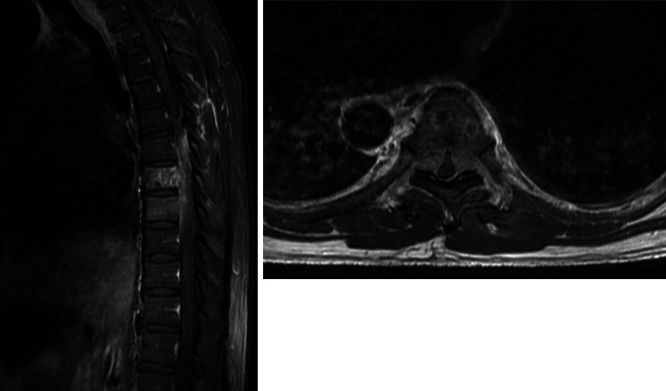
Left: Sagittal postcontrast T1-weighted MRI of the thoracic spine demonstrating T7 and T8 enhancing lesions of the vertebral body with epidural extension. Right: Axial postcontrast T1-weighted MRI of the thoracic spine through the T7 level demonstrating bilateral epidural extension with complete effacement of the cerebrospinal fluid (CSF) spaces and spinal cord compression.