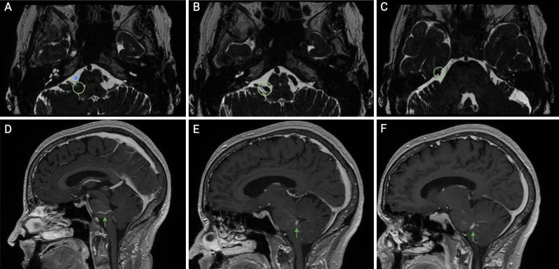
FIG. 1.
Preoperative axial (A–C) and sagittal (D–F) magnetic resonance imaging (MRI) sequences with contrast illustrating a developmental venous anomaly (DVA) (green arrow), the DVA (green circle, A) compressing the facial nerve (blue arrow), and the DVA (green circle, B and C) draining into the superior petrosal sinus.

