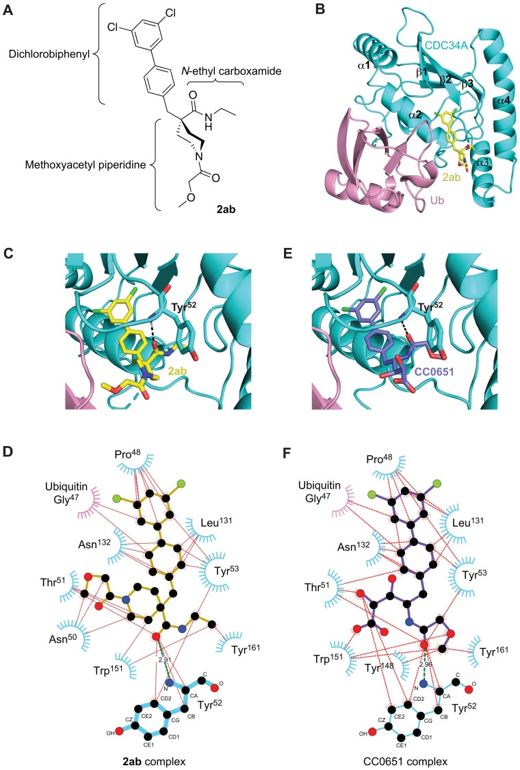
Fig. 7. Structure of 2ab in complex with CDC34A and ubiquitin.
(A) Chemical structure of 2ab drawn to match the orientation depicted in (B). (B) Ribbon representation of CDC34A (cyan) and ubiquitin (pink) with 2ab (carbon atoms in yellow, oxygen in red, nitrogen in blue, and chlorine in green). (C) Zoomed-in view of 2ab in the CDC34A-ubiquitin binding pocket (carbon atoms shown in yellow). See fig. S8A for a stereo view with side-chain details. (D) Ligplot representation of 2ab interactions in the CDC34A-ubiquitin binding pocket. The amide backbone of Tyr52 from CDC34A makes a hydrogen bond with a carbonyl of 2ab (green line) in addition to a network of hydrophobic interactions (red lines). Phe28, Ile45, Phe58, Phe77, Met81, and Ile128 of CDC34A make minor contributions but are not shown for clarity. (E) Zoomed-in view of CC0651 in the CDC34A-ubiquitin binding pocket (carbon atoms shown in purple). See fig. S8B for a stereo view with side-chain details. (F) Ligplot representation of CC0651 interactions in the CDC34A-ubiquitin binding pocket.

