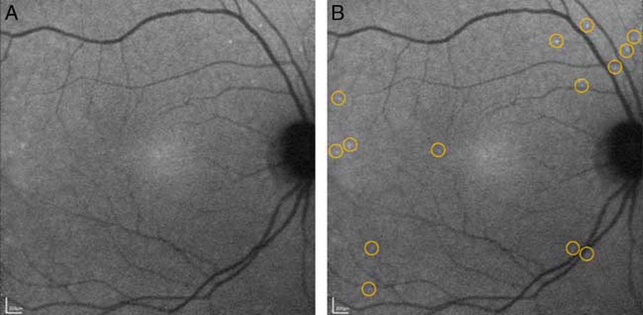
Figure 1.

Detection of apoptotic retinal cells counts in a glaucoma patient showing single cell apoptosis in the retina. 0.4 mg of fluorescently conjugated annexin V was injected intravenously and retinal images were obtained at 240 minutes. Unmarked (A) and marked (B) annexin-positive spots are shown with yellow rings highlighting individual spots. Adapted from Cordeiro et al.30 Adaptations are themselves works protected by copyright. So in order to publish this adaptation, authorization must be obtained both from the owner of the copyright in the original work and from the owner of copyright in the translation or adaptation.
