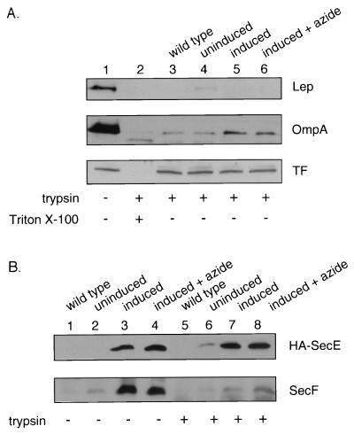
FIG. 1.
The inner membrane remains intact in OMP cells. (A) The levels of the periplasmically oriented marker proteins leader peptidase (Lep) and outer membrane protein A (OmpA) and of the cytoplasmically localized trigger factor (TF) were determined by immunoblotting of OMP cells in the absence (lane 1) or presence (lane 2) of 1.0 mg of trypsin per ml and 1.0% Triton X-100 (Sigma). OMP cells were prepared (11) from BL21 wild-type cells (lane 3), uninduced (lane 4) and induced (lane 5) SecYEG-overproducing cells (12) (induced at an optical density at 600 nm of 0.5 with 1.5% arabinose for 2 h), and induced cells grown in the presence of 20 mM azide for the last 15 min (lane 6). Cells were harvested (5,000 rpm, 5 min, 4°C), resuspended in an equal weight of 10% sucrose–50 mM Tris-Cl (pH 7.5), and frozen in liquid nitrogen. Frozen cells were thawed, collected by centrifugation (5,000 rpm, 5 min, 4°C), and resuspended in 100 μl of 20% sucrose–10 mM EDTA–30 mM Tris-Cl (pH 8.1) for 60 min on ice. Cells were treated with 1.0 mg of trypsin per ml for 60 min on ice. The samples were then precipitated with 15% trichloroacetic acid and acetone washed, and sodium dodecyl sulfate-polyacrylamide gel electrophoresis (SDS-PAGE) was performed with 15% polyacrylamide gels (12). For immunoblotting, proteins separated by SDS-PAGE were electrophoretically transferred to nitrocellulose (Bio-Rad, Hercules, Calif.) with a Genie electrophoretic blotter (Idea Scientific, Minneapolis, Minn.) at 250 mA for 1.25 h. (B) OMP cells were prepared from BL21 wild-type cells (lanes 1 and 5), uninduced (lanes 2 and 6) and induced (lanes 3 and 7) SecYEGDFyajC-overproducing cells, and induced cells grown in the presence of 20 mM azide (lanes 4 and 8). The level of SecE and SecF proteins was determined by immunoblotting of OMP cells incubated in the absence (lanes 1 to 4) or presence (lanes 5 to 8) of 1.0 mg of trypsin per ml. HA, hemagglutinin.

