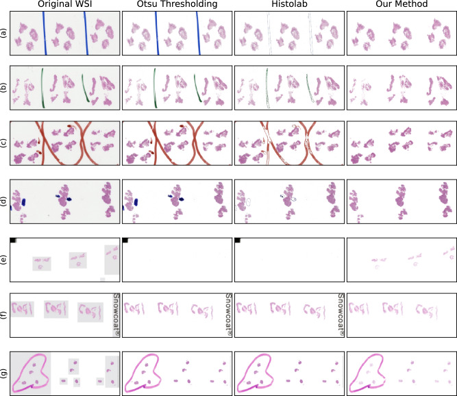
Figure 3.
Seven WSIs of H&E-stained biopsies containing artefacts of a wide range of types and colours. The aim was to segment the tissue without including background and artefacts. First Column: The original WSI. Second Column: The tissue segmentation provided by applying Otsu thresholding to the luminance of the WSIs placed on a white background. Otsu thresholding failed to reject a single artefact and failed to segment the tissue in (e). Third column: the tissue segmentation from Histolab tissue thresholding and pen filters. While there was partial pen mark removal in (a–d), the pen marks were not fully removed in any image, no tissue was segmented in (e), no pen marks were removed in (g). Forth column: the tissue segmentation from our method placed on a white background. Our method successfully segmented all tissue and rejected all background and artefacts except the pen marks in (g). Our method failed to reject the pen marks in (g) because the pen is the same colour as the eosin.

