Figure 7.
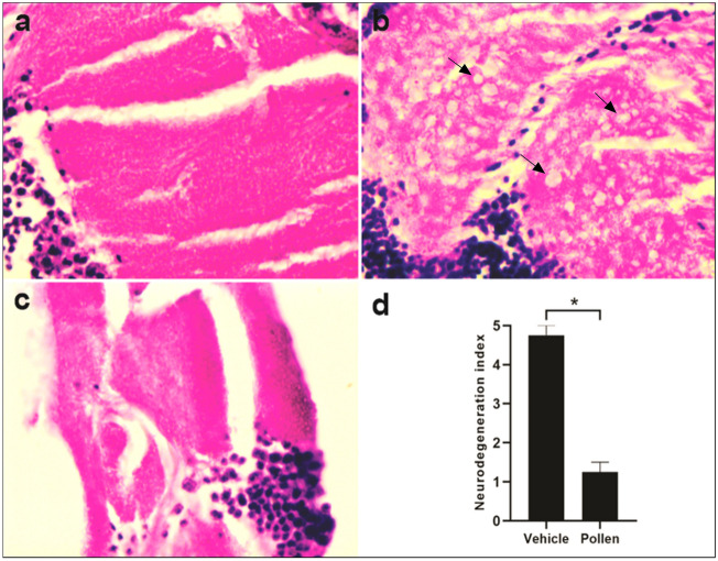
Histology of the brain of AD-like model fly treated with methanolic pollen extract. Representative 3-μm paraffin sections brain of flies 15-days after hatching. Illustrative images of histopathologic analysis of (a) elav-Gal4, magnification of ×100; (b) AD-like treated with vehicle (tween), ×100—arrows indicate vacuolar lesions, and (c) AD-like flies treated with pollen, ×100. (d) Neurodegeneration index of AD-like flies treated with vehicle (tween) and pollen based on histopathology analysis, according to vacuolar lesions. 0 indicates no lesions, and 5 indicates neurodegenerative phenotype (n = 3, at 15 days after treatment). Data are shown as the mean ± SEM. The statistical significance is indicated as * for P < 0.05 (Mann Whitney test).

