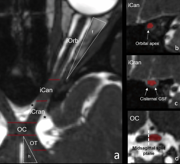
Figure 1.
Illustration of the aVP subdivision boundaries applied for segmentation, as seen in one subject on the CISS images. The left panel shows a curved reconstruction of the aVP along its longitudinal axis, with red bars marking the boundaries (a). The iOrb/iCan boundary is located at the end of the intraorbital fat (f) and corresponds to the orbital apex at the level of the annulus of Zinn envisioned in (b). The transition from iCan to iCran is marked by the cisternal CSF (*) surrounding the nerve as seen in (c). The OC is the segment where the left and right aVPs merge perpendicular to the midsagittal plane (dashed line in d). The hypothalamus (h), representing the medial OT boundary, is highlighted in (a).

