
Fig. 6.
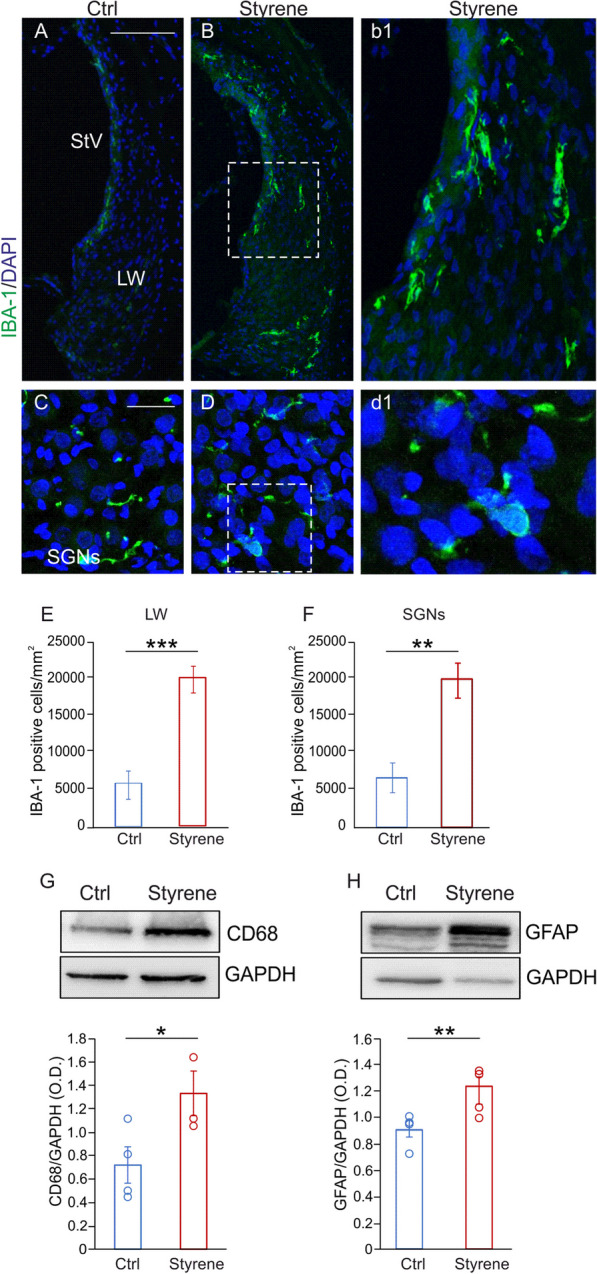
Macrophages play a role in ototoxic damage induced by styrene. A–D Images of cochlear sections showing high magnifications of the lateral wall (LW) with stria vascularis (SV; A, B) and spiral ganglion neurons (SGNs; C, D) stained with IBA-1, (green fluorescence) and DAPI (blue fluorescence). High magnifications show a strong IBA-1 fluorescence in the LW (b1) and SGNs (d1) in styrene cochlear samples. Scale bar: A, B 100 μm; C, D 50 μm. E, F Histograms showing IBA1-positive cell density in the LW (E) and in SGNs (F). Data are expressed as mean ± SEM and are representative of three independent experiments from three animals/group. G, H Representative western blots showing high levels of CD68 and GFAP in cochlear lysates in Styrene compared to Ctrl groups. Histograms (means ± SEM) show the optical density values (CD68 n = 3 cochleae for each group, p = 0.03; Student’s t test; GFAP n = 4 cochlea for each group; Student’s t test, p = 0.009) normalized to the corresponding total protein amount (GAPDH). Asterisks show statistical significance (*p < 0.05; **p < 0.01; ***p < 0.001)