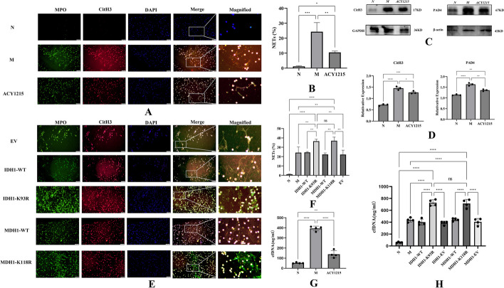
Fig. 3.
IDH1/MDH1 deacetylation enhanced NETs formation. A dHL-60 cells were stimulated with PMA in vitro, left untreated/treated with ACY1215, and immunofluorescently stained with MPO (green), CitH3 (red), and DAPI (blue) to detect the formation of NETs; the scale bar is 50 μm. B NETs and dHL-60 cells were counted after immunofluorescence staining with MPO and CitH3. The percentage of NETs was calculated as the average of five fields normalized to the total number of dHL-60 cells. C In dHL-60 cells, the protein levels of CitH3 and PAD4 were measured by western blotting. D In dHL-60 cells, the protein levels of CitH3 and PAD4 were analyzed by ImageLab. E In dHL-60 cells, with plasmid mutations at K118 of MDH1 and K93 of IDH1, cells were stimulated with PMA in vitro, and immunofluorescence staining with MPO (green), CitH3 (red), and DAPI (blue) was performed to detect the formation of NETs; the scale bar is 50 μm. F NETs and dHL-60 were counted after immunofluorescence staining with MPO and CitH3 to calculate the proportion of dHL-60 releasing NETs. G NETs were quantified by a Quant-iT™ PicoGreen® dsDNA kit. H NETs were quantified by a Quant-iT™ PicoGreen® dsDNA kit. N:dHL-60; M: dHL-60 + PMA; ACY1215: dHL-60 + PMA + ACY1215;IDH1-WT: IDH1-wild-type + PMA; IDH1-K93R:IDH1-K93R + PMA; IDH1-EV: IDH1-Flag + PMA; MDH1-WT: MDH1-wild-type + PMA; MDH1-K118R:MDH1-K118R + PMA; MDH1-EV: MDH1-HA + PMA, NETs Neutrophil extracellular traps, cfDNA Cell-free DNA; MPO Myeloperoxidase, CitH3 Citrullinated histone H3, DAPI 4′,6-diamidino-2-phenylindole, PAD4 peptidylarginine deiminase 4.The histograms show the means ± SD. ns no significance, *P < 0.05, **P < 0.01, ***P < 0.001, and ****P < 0.0001

