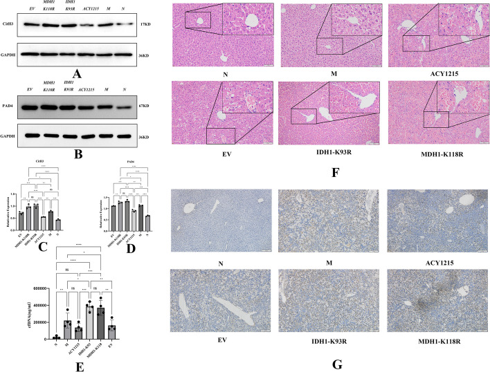
Fig. 5.
IDH1/MDH deacetylation promotes acute liver failure by regulating NETosis. A, C The expression of hepatic CitH3 was measured by western blotting and analyzed by ImageLab. B, D The expression of hepatic PAD4 was measured by Western blotting and analyzed by ImageLab. E NETs were quantified by a Quant-iT™ PicoGreen® dsDNA kit. F HE staining of mouse liver tissue under different treatments; the scale bar is 100 μm. G Cell apoptosis was detected by the TUNEL method in the liver tissue of mice under different treatments; the scale bar is 100 μm. N: liver samples injected with normal saline; M: ALF model, liver samples injected with LPS/d-gal; ACY1215: LPS/d-gal + ACY1215; IDHI-K93R: IDHI-K93R adenovirus-infected mice + LPS/d-gal; MDHI-K118R: MDHI-K118R adenovirus-infected mice + LPS/d-gal group; EV: empty adenovirus-infected mice + LPS/d-gal, CitH3 citrullinated histone H3, PAD4 peptidylarginine deiminase 4, cfDNA Cell-free DNA. The histograms show the means ± SD. ns no significance, *P < 0.05, **P < 0.01, ***P < 0.001, and ****P < 0.0001. *P < 0.05, **P < 0.01, ***P < 0.001, and ****P < 0.0001

