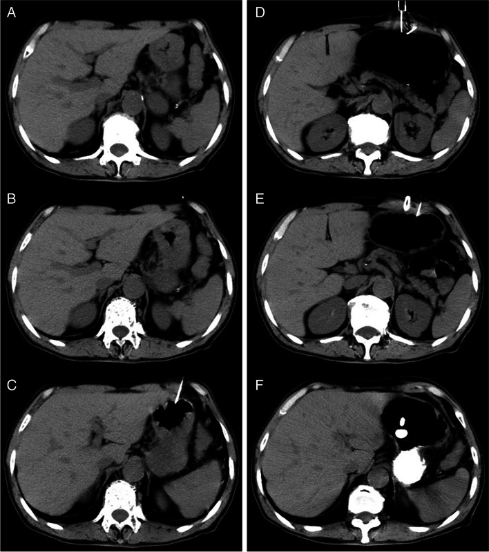
Fig. 1.
A 56-year old man who cannot underwent the gastrostomy under DSA guided due to the esophagus complete obstruction was referred to our department to undergo a CT guided percutaneous gastrostomy for long-term intragastric feeding. A A prepared abdominal CT scan was performed to localize the stomach and design the puncture pathway. B A metallic marker was placed on the skin surface to show the puncture pathway. C A 21-gauge Chiba needle was introduced into the gastric lumen and then the air inflation of gastric lumen was performed through the 21-gauge Chiba puncture needle. D After the stomach lumen insufflations finished, puncture procedure was performed with the gastropexy device. The complete gastropexy device was removed, maintaining traction on the wire loop retrieving the suture, which was released and tied in a standard surgical knot, opposing the anterior stomach to the anterior abdominal wall. E After successful gastropexy at both marked points a stab incision of about 5 mm was performed in between to insert the trocar with peel-away sheath carefully under CT guidance. The CT scan showed the trocar with peel-away sheath and the 21 G Chiba needle. F Once correct position was confirmed the trocar was removed and the balloon tube was inserted into the gastric lumen. The CT scan showed the balloon tube filled with 3 ml of contrast medium through an extra valve on the side using a syringe.

