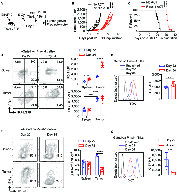
Fig. 3.

IRF4 expression in adoptively transferred antitumor CD8+ T cells in melanoma. Thy1.2+ B6 mice were s.c. injected with 0.2 × 10⁶ B16F10 cells on day 0. The mice were then sub-lethally irradiated and adoptively transferred with 2 × 10⁶ activated Irf4GFP-DTRThy1.1+ Pmel-1 CD8+ T cells (Pmel-1 ACT) or without any T cell transfer (No ACT) on day 3. Tumor growth was monitored, and adoptively transferred Pmel-1 cells were analyzed on days 22 and 34. (A) Schematic of the experimental design. (B and C) Tumor volumes and survival rates of B16F10 tumor-bearing mice in the Pmel-1 ACT (n = 10) and No ACT (n = 7) groups. (D) IRF4.GFP and PD-1 expression of Pmel-1 cells in spleens and tumors of the Pmel-1 ACT group on the indicated days. (E) TOX expression of Pmel-1 TILs on indicated days. (F) Percentage of IFN-γ+TNF-αhigh cells among Pmel-1 T cells in spleens and tumors. (G) Ki-67 expression of Pmel-1 TILs. In B, tumor growth curves (from day 8 to day 22) were compared between the Pmel-1 ACT and No ACT groups using a 2-way ANOVA (mixed-effects model) with the Geisser–Greenhouse correction. In C, survival rates were compared between the Pmel-1 ACT and No ACT groups using a log-rank test. In D to G, the results in bar graphs are presented as mean ± SD (n = 6 to 8), and statistical significance was determined using an unpaired 2-tailed Student’s t test. **P < 0.01, ***P < 0.001, ****P < 0.0001.
