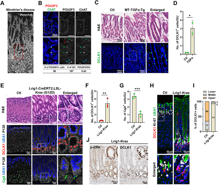
Figure 2.

Expansion of doublecortin‐like kinase 1 (DCLK1)‐positive cells in human Ménétrier's disease and mouse models of foveolar hyperplasia. (A and B) Images of Hoechst (A) and co‐immunostaining for POU2F3 and ChAT (B) in the stomach of patient with Ménétrier's disease. Panel (B) corresponds to the red dotted box area in panel (A). (C and D) Representative images of H&E and immunostaining for DCLK1 in the corpus from control and MT‐transforming growth factor α transgenic (MT‐TGFα‐Tg) mice 2 weeks after induction (C) and quantitation of tuft cells per gland in the stomachs of control (n = 2) and MT‐TFGα‐Tg (n = 2) mice (D). (E) Representative images of H&E or co‐immunostaining for DCLK1, UEA1, P120, or CgA in the corpus from control and Lrig1‐Kras mice at 2 months after induction. Dotted box depicts enlarged area. (F and G) Quantitation of DCLK1‐positive (F) and CgA‐positive cells (G) in control (Ctl, n = 3) and Lrig1‐Kras (n = 3) mice. (H) Co‐immunostaining for DCLK1, Ki‐67, and UEA1. (I) Proportion of tuft cells in the lower, middle, and upper areas of gastric glands. (J) Immunohistochemistry for p‐ERK and DCLK1 in Lrig1‐Kras mice at 2 months after induction. No., number; GU, gastric unit. Mean ± SD. Unpaired Student's t‐test or one‐way ANOVA with Tukey's multiple comparisons test. *p < 0.05, **p < 0.01, ***p < 0.001, ****p < 0.0001.
