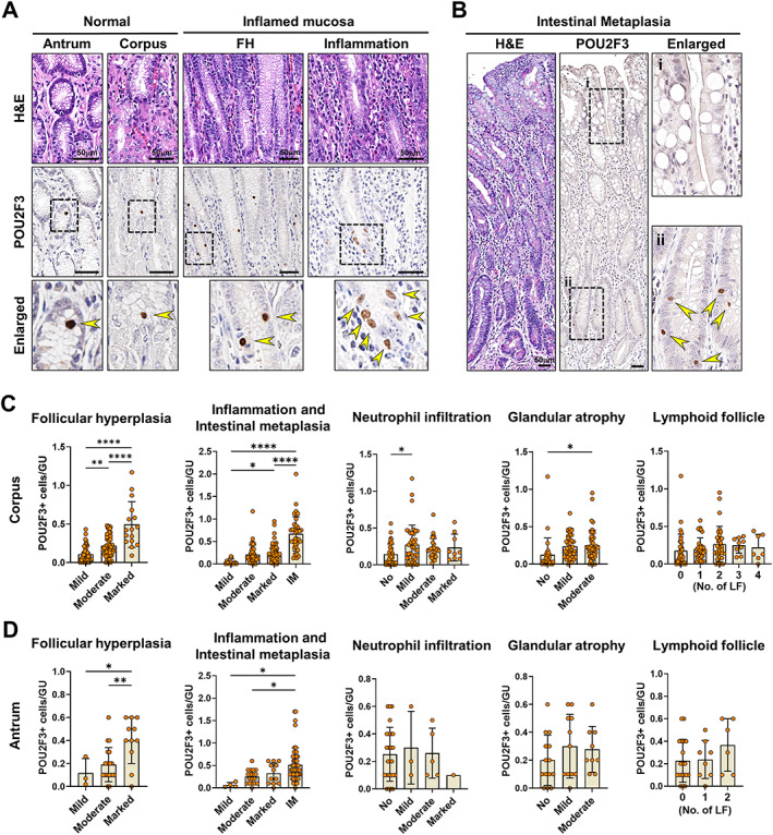
Figure 3.

Associations between tuft cells and pathological features of human gastric lesions. (A) Representative images of H&E and immunostaining for POU2F3 in the normal antrum and corpus and gastric mucosa with foveolar hyperplasia (FH) or inflammation. (B) H&E and immunostaining for POU2F3 in the IM of stomach. (C and D) Quantitation of POU2F3‐positive cells per gastric unit according to the pathological features such as FH, chronic inflammation, activity, glandular atrophy, IM, and lymphoid follicles (LFs) in the antrum (C) and corpus (D). GU, gastric unit. Mean ± SD. One‐way ANOVA with Tukey's multiple comparisons test. *p < 0.05, **p < 0.01, ****p < 0.0001.
