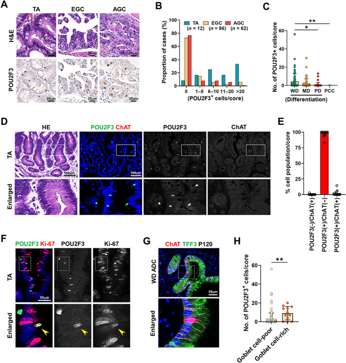
Figure 5.

Characteristics of tuft cells in human gastric adenomas and cancers. (A) Representative images of H&E and immunostaining for POU2F3 in the TAs, EGC, and AGC. (B) Percentages of cases with various numbers of POU2F3‐positive cells in the TA (n = 8), EGC (n = 86), and AGC (n = 62). (C) Quantitation of POU2F3‐positive cells per core according to differentiation of cancer glands (WD, well differentiated, n = 73; MD, moderately differentiated, n = 87; PD, poorly differentiated, n = 43; PCC, poorly cohesive carcinoma, n = 33). (D and E) H&E and co‐immunostaining for POU2F3 and ChAT in TAs (D) and proportions of cells according to POU2F3 and ChAT positivity (E). (F) Co‐immunostaining for POU2F3 and Ki‐67 in TA. (G and H) Co‐immunostaining for ChAT, TFF3, and P120 in WD adenocarcinoma (G) and quantitation of POU2F3‐positive cells per core in goblet cell‐poor or goblet cell‐rich gastric cancers (H). No., number. Mean ± SD. Unpaired Student's t‐test or one‐way ANOVA with Tukey's multiple comparisons test. *p < 0.05, **p < 0.01.
