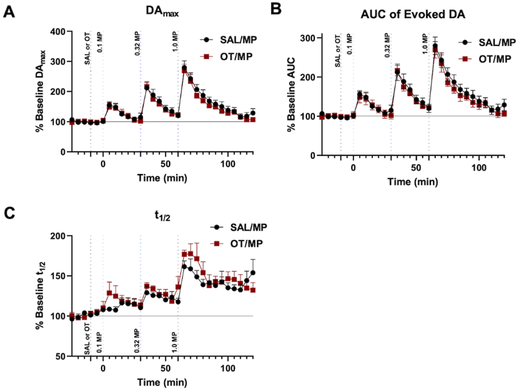
Figure 4: Oxytocin (OT) administration does not alter methylphenidate (MP)-induced increases in phasic nucleus accumbens shell (NAS) dopamine (DA).

Analysis of fast scan cyclic voltammetry (FSCV) results of evoked NAS DA at baseline/control, 10 min after saline or OT (2mg/kg; i.p.) and 10 min after increasing doses of methylphenidate (MP) (0.1, 0.32, 1.0 mg/kg; i.v.). Data are shown as the average of each animal’s results normalized to their baseline for (A) Maximum evoked DA (DAmax), (B) area under the curve (AUC), (C) t1/2 throughout the experimental paradigm and error bars indicate SEM (n=6 rats for each).
