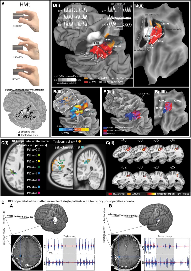
Figure 4.
Spatial matching between DES and SVR-LSM results. (A) Hand manipulation task (HMt) and sampling of parietal stimulation from Fornia et al.21 [B(i, ii)] Co-localization between HMt probability density estimation (effective areas in white and ineffective areas in black), praxis cluster (red) and prehension-praxis common region (orange). The upper part of [B(i)] shows examples of EMG interference patterns evoked by parietal DES of phAIP and PF. [B(iii)] HMt probability maps showing the parietal region associated with different EMG-interference patterns (task-clumsy versus task-arrest) regardless of ineffective sites. [B(iiii)] Co-localization between HMt task-arrest and clumsy pattern probability with meaningful (blue), meaningless (red) gestures CFWER (covariate ARAT) and posterior parietal regions (phAIP, DIPSA, DIPSM, PF). [C(i)] Anatomical localization of effective sites recorded within the parietal white matter. [C(ii)] Probability density estimation of HMt effective sites within the white matter and their co-localization with praxis CFWER (covariate ARAT). (D) Example of two patients showing transient postoperative apraxia: (A) the effective site was located in the white matter below the AIP and evoked a task-arrest pattern; (B) the effective sites were located in the white matter below the PF and evoked task-clumsy patterns. ARAT = Action Research Arm Test; CFWER = cluster-level family-wise error correction; DES = direct electrical stimulation; DIPSA = dorso-anterior intraparietal sulcus; DIPSM = dorso-medial intraparietal sulcus; phAIP = putative homologue of anterior intraparietal, AIP; SVR-LSM = support vector regression lesion symptom mapping.

