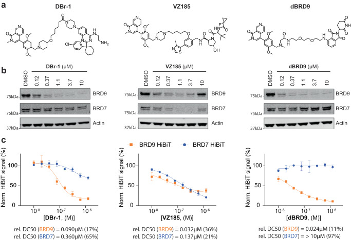
Fig. 3. Comparison of different BRD9 PROTACs.
a Compound structures of the DCAF1-BRD9 PROTAC DBr-1, the VHL-BRD9 PROTAC VZ185 and the CRBN-BRD9 PROTAC dBRD9. b Immunoblot analysis of HEK293 BRD9-HiBiT/FF/CAS9 cells treated for 2 h with DBr-1, VZ185, and dBRD9 at various doses. c BRD9-HiBiT and BRD7-HiBiT signal detection of samples treated for 2 h with DBr-1, VZ185, and dBRD9 at various doses, DC50 values and maximal observed degradation are shown below. Data represents mean ± standard deviation from n = 3 replicates. Source data are provided as a Source Data file.

